Technology
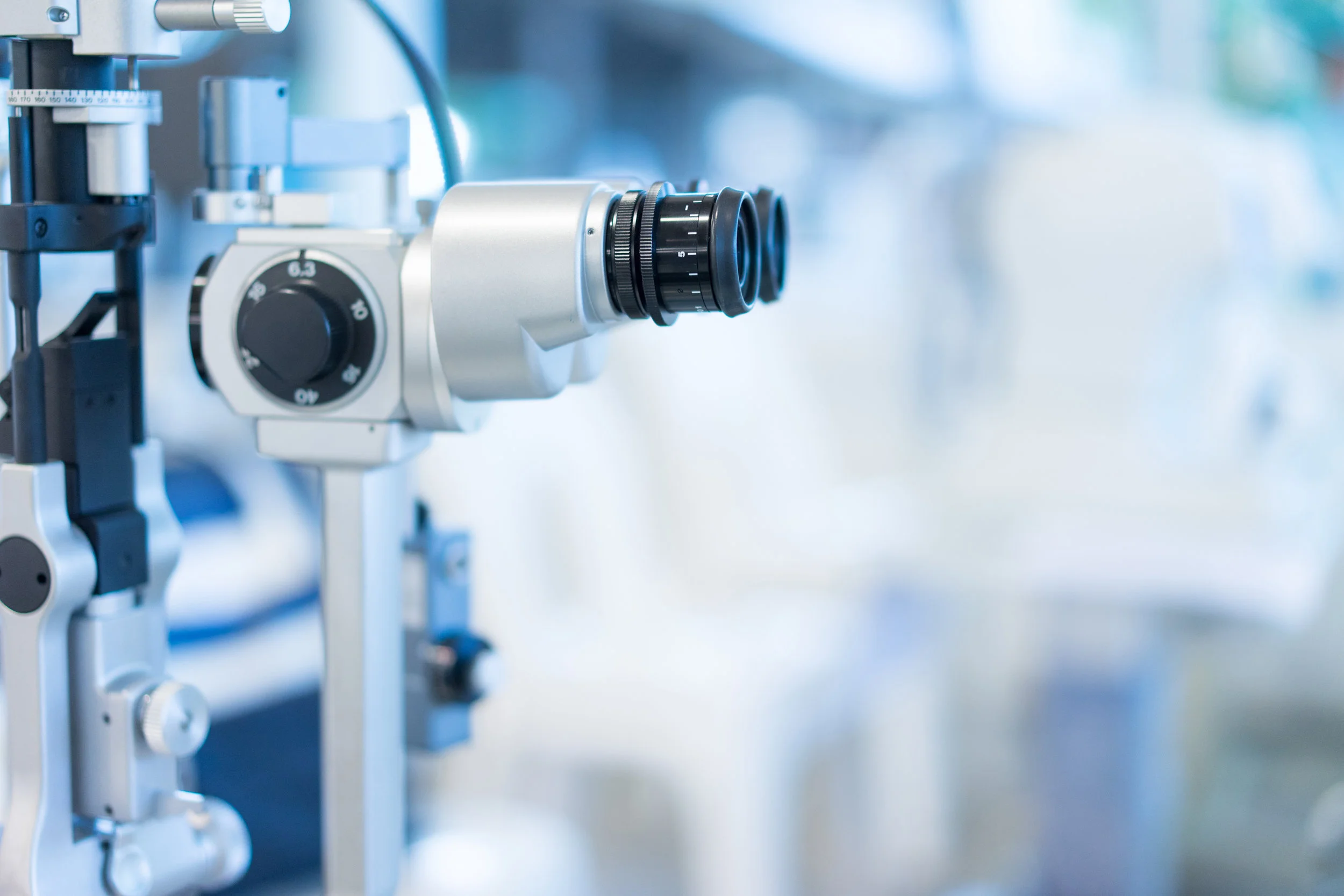
Medicine is ever progressing. The way we can view the eye and detect the early levels of diagnostic changes now – is nothing less than miraculous. We can test pupillary functions at speeds to the millisecond, retinal thicknesses to the micron and observe, microscopically, to such levels as to see red blood cells tumbling in the arteries. Our progressive approach to examination leads the profession in enhanced neuro-electrical testing, imaging and functional evaluation. At Advanced Clinical Eye Care of Maine we strive to bring you the best instrumentation to allow our talented doctors to evaluate and diagnose at levels unobtainable by conventional methods.
Our doctors and staff train constantly. We attend lectures and seminars, wet labs and technical instruction to learn all that we are able so we may bring this knowledge to you the second it becomes available. Yet, we recognize the high costs of health care and therefore have some of the lowest fees in the area to allow our patients access to this high tech care that allows us to provide the best eye care possible.
Optical Coherence Tomography
Optical Coherence Tomography is a device that uses light waves to create images of the inside of the eye, much like an ultrasound uses sound waves to image the inside of the body. This allows the doctor to analyze each and every layer of the eye in cross section and in layers like a topographical map. With this information we can quantify the thickness of tissues and their overall heath. This is now essential equipment in a complete retinal evaluation for disorders such as; tears, fluid leakage, tumors, and macular degeneration. This test is becoming the gold standard in diagnosis of Glaucoma.
Anterior Segment Imaging
The front chamber of the eye is the section from the front of the lens forward and includes the Iris, pupil and cornea. This new imaging technology allows us to view areas otherwise hidden behind the iris and unseen by standard microscopy. This imaging allows us to follow and monitor; corneal issues, iris cysts and tumors, evaluate the drainage of fluid in glaucoma, and integrity of the eye in post-operative cataract surgery.
Corneal topography
This is a non-invasive method to map the cornea much like a topographical map in order to evaluate the surface and monitor changes in the surface. This device makes it possible to follow things like; corneal transplants, scarring, surface disease, and keratoconus. This technology also allows for the precise fitting of contact lenses, especially over compromised surface and in high refractive errors and astigmatism.
Frequency Doubling Field Testing
This equipment allows us to evaluate the integrity of both the retina and the optic nerves by testing the threshold of light the system can perceive and react to. It is more specific in allowing the separation of the different layers of the pathways than convention field testing alone. This testing is a first step in screening for glaucoma and in diabetic patients for retinopathy.
Optos
The Optos imaging system is an ultra-wide field imaging system that can view and capture images of the retina like nothing else in its class. This device allows the doctor, to not only analyze the health and structure of these tissues, but enables them to display and show the patient their own images to better help them understand their specific condition.
Lipiscan
Many dry eye patients suffer from decreased secretions from the Meibomian glands. These glands are embedded in the upper and lower lids of each eye. The secretions from these glands, creates the oily outer layer of the tears and helps hold the tear up and helps prevent evaporation. The Lipiscan is a revolutionary new tool to image this glans and aids the doctor in assessing the viability of these glands. It is a critical tool in the diagnosis and treatment of Dry Eye disease.
Lipiflow and TearCare: Advanced Treatment for Meibomian Gland Dysfunction
Thermal pulsation treatments, such as LipiFlow and TearCare, are designed to address meibomian gland dysfunction (MGD)—a common cause of dry eye. These devices gently warm the eyelids to a therapeutic temperature (around 105°F) to soften the oils in the glands. Once the oil is loosened, the glands are carefully massaged to clear blockages and restore healthy oil flow to the tear film. Clinical studies have shown these treatments can significantly improve gland function and provide lasting relief from dry eye symptoms.


